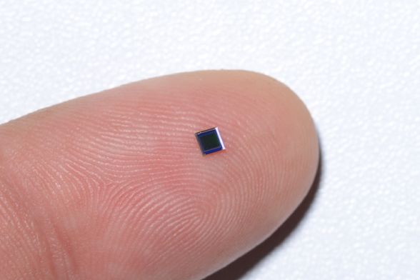
IN Brief:
- Sony's IMX742 is a 1 mm-square CMOS sensor developed for cameras used in single-use endoscopes.
- The sensor provides 720 × 720 imaging at up to 60 fps and carries video and control signals through a compact six-pin connection.
- Samples are scheduled for November 2026; five-metre digital transmission was demonstrated under specified test conditions, not qualified in a finished medical device.
Sony Semiconductor Solutions has developed the IMX742, a CMOS image sensor measuring 1 mm square for cameras used in single-use medical endoscopes. The device combines 720 × 720 effective pixels, digital image output, and a compact six-pin package, with sample shipments planned for November 2026.
Sony is targeting applications such as bronchoscopes and ureteroscopes, where the space available at an instrument’s distal tip restricts the dimensions of the sensor, optics, illumination, and electrical connections. The image sensor is a component for equipment manufacturers, rather than a complete camera or an approved medical endoscope.
The back-illuminated device has a diagonal active image size of approximately 1.02 mm, described as Type 1/17.7, and uses 1.0 µm pixels. Its 720 × 720 pixel arrangement corresponds to 518,400 effective pixels, which Sony describes as approximately 0.51 megapixels under its published specification method. It supports operation at either 30 or 60 frames per second.
Reducing the dimensions of a sensor involves compromises in the area available for each light-sensitive pixel and for the circuits that convert the resulting signals. A compact endoscope camera also has limited room for illumination equipment, making image noise and the available light particularly important in the overall system design. Sony has miniaturised peripheral circuits, including the analogue-to-digital converter, while retaining the specified pixel geometry.
Digitising the image signal on the sensor changes how information travels along the endoscope. An analogue camera sends a continuously varying electrical signal through the cable, leaving the receiving electronics to convert it into digital image data. Signal degradation and electrical interference along the connection can therefore affect the received image. The IMX742 performs conversion at the sensor, so the cable carries digital data rather than the analogue pixel signal.
Sony has demonstrated digital transmission through a five-metre cable using AWG44 wire in its own evaluation environment. The company describes the combination of digital output and this transmission length as an industry first among medical endoscope CMOS image sensors, according to its research. Both the priority claim and the transmission result belong to Sony: the result is not a guarantee that every five-metre cable or finished endoscope assembly will achieve identical performance.
Cable geometry, connectors, electromagnetic interference, and the receiving circuitry can all affect signal integrity. In a medical imaging instrument, the cable must also meet mechanical requirements associated with bending, handling, and routing through a narrow insertion tube. These factors require validation at equipment level, even when the electrical connection works in the component manufacturer’s laboratory.
The IMX742 uses proprietary bidirectional communication to carry control and video information through the same cable arrangement. This reduces the number of separate signal paths that have to reach the sensor and allows a six-pin wafer-level chip-scale package. Camera designers can then allocate limited space to the optics, light source, and mechanical assembly, although the finished module will necessarily be larger than the sensor chip alone.
Image quality depends on more than the number of pixels. Exposure settings, lens design, illumination geometry, and downstream processing affect the clarity of tissue structures, especially where the camera operates in relatively dark spaces. Sony reports less graininess and fewer vertical image artefacts than a conventional analogue-output arrangement in its comparison images. Those illustrations are company evaluations, not clinical performance measurements.
The maximum 60 fps rate can improve the continuity of displayed movement, but usable image quality at that frame rate depends on sufficient light and suitable exposure. The device uses a rolling shutter with global reset functionality; this should not be interpreted as a conventional full global-shutter sensor. Its Bayer colour filter requires subsequent processing to produce colour images.
Electrical integration also requires three stated supply rails of 2.7 V, 1.2 V, and -1.2 V. The camera manufacturer must provide appropriate regulation, sequencing, and protection while managing heat and electrical noise near the sensor. Sony’s published specification defines the component’s interfaces and characteristics, but the complete camera system will determine the practical operating envelope.
Single-use endoscopes are being developed partly to reduce the burden of cleaning and reprocessing reusable instruments and to address associated infection-control concerns. Those potential benefits cannot be inferred from the sensor on its own. Sony explicitly states that the IMX742 is not a medical device and has not been evaluated or approved for medical applications; any finished instrument must undergo its own validation and regulatory assessment.
Sony plans to begin sample shipments in November 2026, providing developers with hardware to evaluate the optical, electrical, and mechanical integration. No timetable for certified endoscope products using the IMX742 has been disclosed. The immediate advance is a digitally connected sensor whose small dimensions allow equipment developers to investigate narrower imaging assemblies without assuming that component-level tests establish device-level performance.